CARBOHYDRATE-CHEMISTRY

Research

Image of Doctor Andreas Franz  Image of Franz research group showing five students outside the chemistry building on South campus, spring 2025

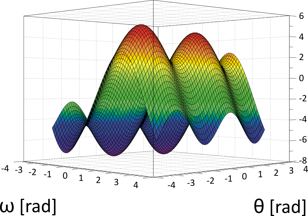
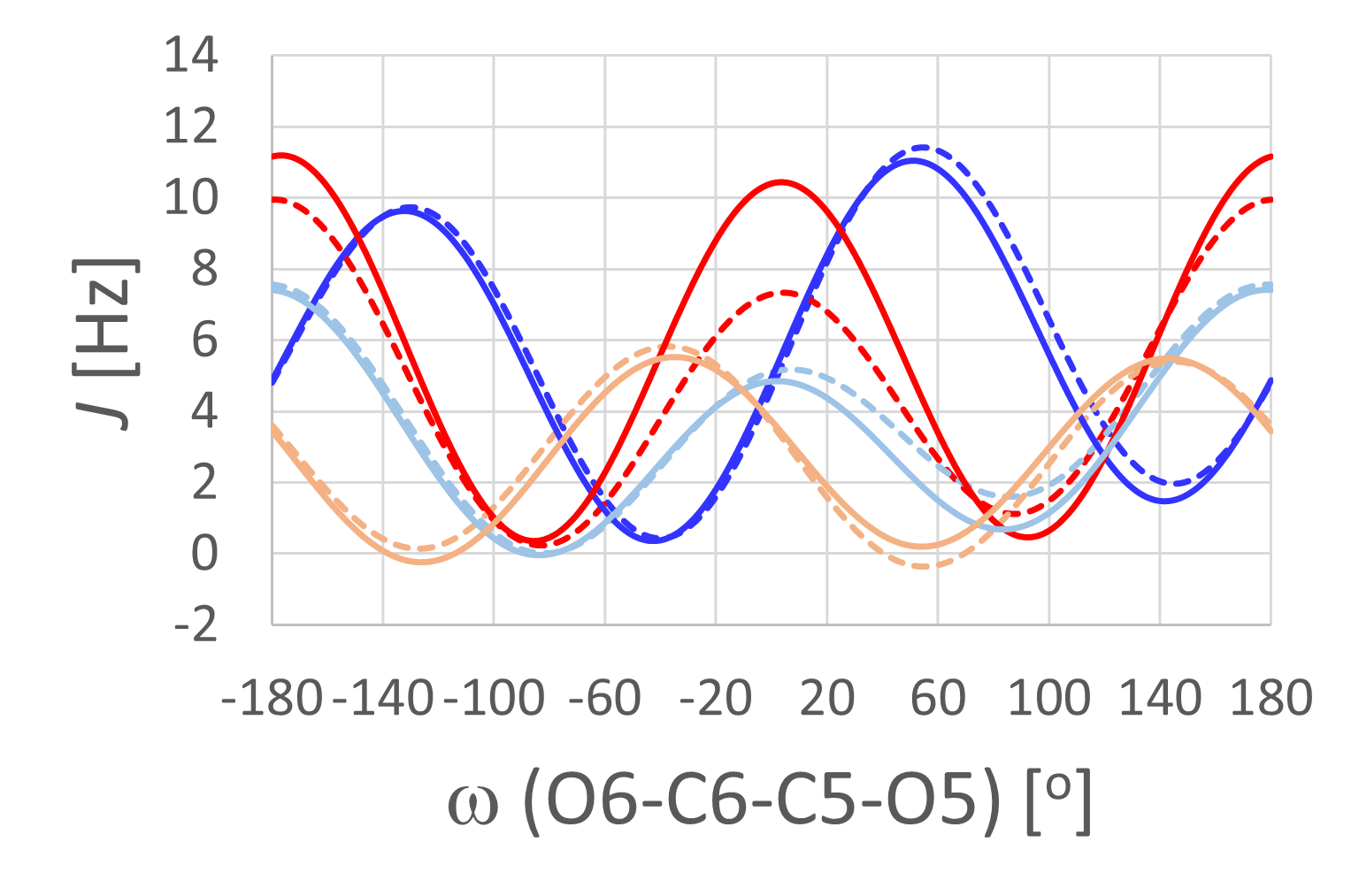
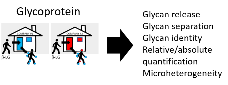
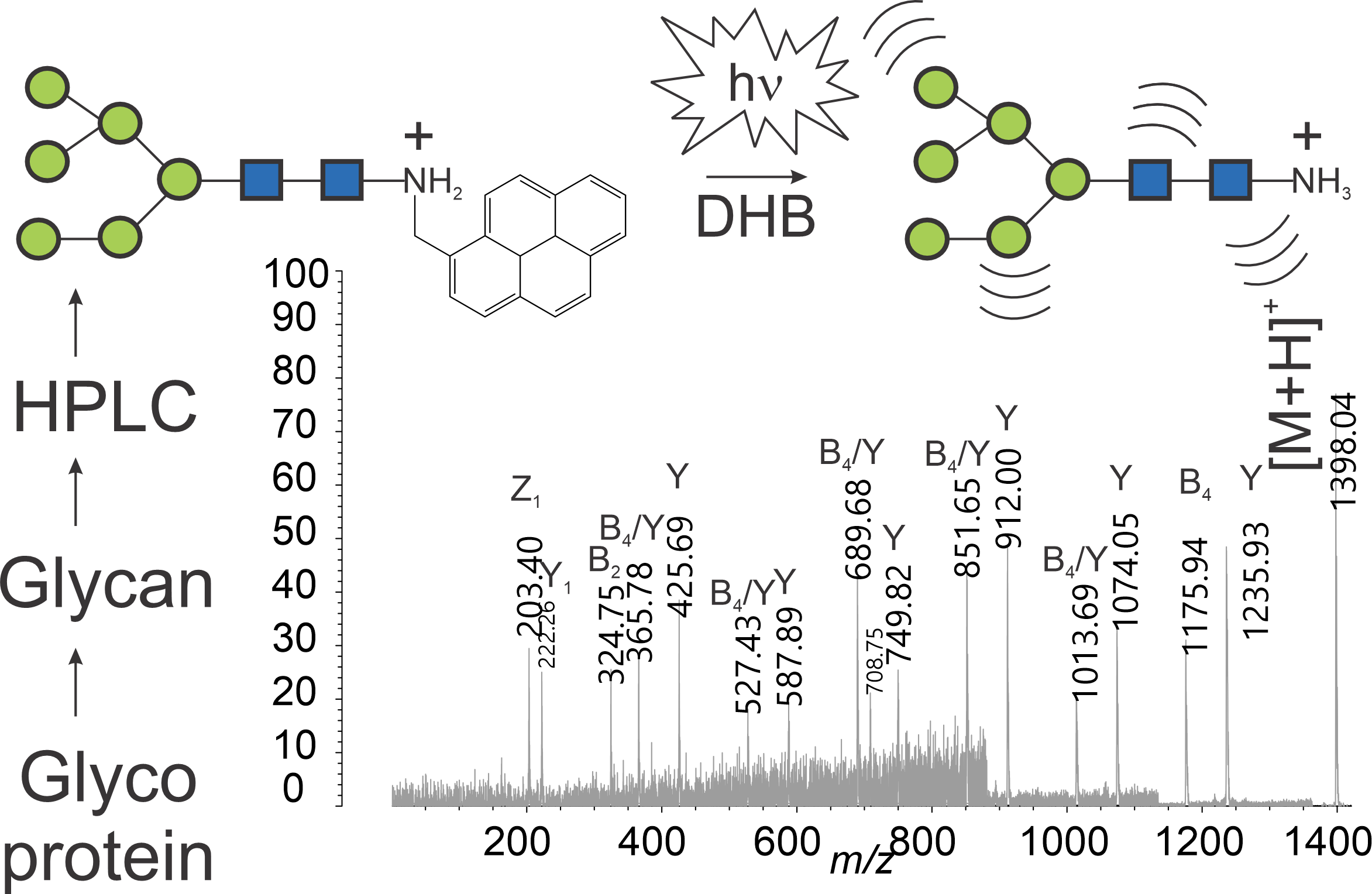
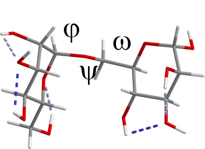
Solution Conformation of Carbohydrates by NMR: Compounds with hydroxyl clusters, such as carbohydrates, have potentially biologically interesting properties. They have been implicated in a multitude of biochemical processes that are central to life on earth. The structural diversity among carbohydrates and - as a consequence - their potential to "code" for biochemical information has no parallel. It is, therefore, striking that nature only uses a comparatively small number (~ a few thousand) of structures to make biochemistry on this planet possible. The accurate description of carbohydrate conformation in aqueous solution is critical before function can be understood. The solution conformation is complicated by many different factors. We use NMR spectroscopy and theoretical methods to develop an acceptable quantitiative description.

Hydrogen Bonding in Carbohydrates: Exchangeable protons contribute to hydrogen bonding during biochemical recognition events. However, exchangeable protons are difficult to detect by NMR spectroscopy. We use H/D-exchange techniques and the resulting chemical isotope shift effects at variable temperatures to detect evidence for transient hydrogen bonding.

Oligosaccharide Tags: Glycans play important roles in inflammation, fertilization, immunocompatibility, and cell growth. The inherent difficulty in detecting oligosaccharides released from glycoproteins due to  the absence of chromophores has been addressed in the past by introducing UV-active or fluorescent labels into the sugar prior to chromatographic separation. We develop and evaluate new labeling reagents for oligosaccharides. The glycan derivatives are purified and studied by MALDI-TOF mass spectrometry.

Collaborative projects with Dr. George Pantouris and Skylar Carlson

Image showing a stick molecular model of isomaltose including 
torsion angle labels of phi, psi, and omega along the alpha 1 6 glycosidic linkage

Two proton NMR spectra of isomaltose in water. The bottom trace
shows the spectrum recorded in d two o at twohundred ninety eight Kelvin room temperature. The inset between six and seven p p m shows no signals from hydroxyl 
group protons. The top trace shows the spectrum recorded in ninety nine percent h 2 o at decreased temperature of twohundred sixty eight Kelvin. The inset between 
six and seven p p m shows clear signals for hydroxyl protons

Three dimensional plot showing a Karplus hypersurface for J coupling as a function of 
torsion angle omega and theta. High J-values are shown in red and yellow, medium J-values are shown in green, and small or negative J values are shown in blue

Plot showing multiple Karplus equations for J coupling as a function of the torsion 
angle omega defined by O six, C six, C five, and O five


Image showing the word glycoprotein with two houses below it. One house is red and the 
other house is blue. A cartoon stick figure walks into the blue house and comes out with blue dots on its body. Another cartoon stick figure walks into the red 
house next door and comes out with both blue and red dots on its body. An arrow points to a list of words to the right of the two houses and stick figures. The list 
is of the words glcyan release, glycan separation, glycan identity, relative slash absolute quantification, and microheterogeneity


Image showing a flow chart including text and symbolic oligosaccharide structures above 
a MALDI time of flight mass spectrum. The flow chart starts with the word glycoprotein, followed by glycan, followed by H P L C, followed by the oligosaccharide 
structure of the high mannose six glycan with six green circles and two blue squares. The structure also shows a covalent pyrene methyl amine tag and a positive charge 
on the nitrogen atom. The next flow chart entry shows a flash of light cartoon and h times nu along with D H B followed by the high mannose six glycan structure lacking 
the pyrene label. The structure appears to vibrate. Below is the mass spectrum showing many peaks at different m over z values


INTERESTED TO JOIN?

Graduate Students

Undergraduate Students


Homepage| Research| Publications| Coworkers | Chemistry Resources| Courses| FAQ

College of the Pacific | Pharmaceutical Sciences and Chemistry

Updated: Apr. 2025